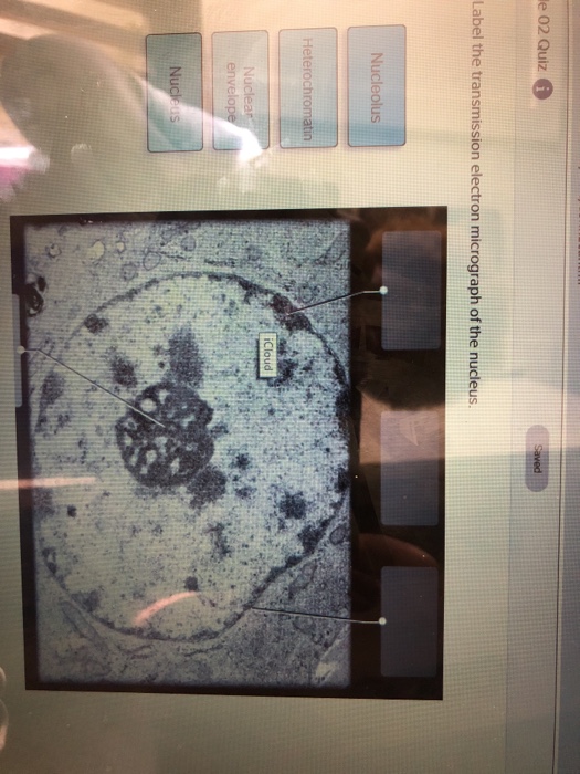
33 Label The Transmission Electron Micrograph Of The Nucleus
Wide collections of all kinds of labels pictures online. The electron micrograph of mitochondria.
 Localization Of Phosphorylated Connexin 43 Using Serial Section
Label the transmission electron micrograph of the nucleus b978141602255850022x gr1.

Label the transmission electron micrograph of the nucleus. A form of electron microscope in which an image is derived from electrons that have passed through the specimen in particular one in which the whole image is formed at once rather than by scanning. At the center of the image is a large structure called the nucleus which is a membrane limited compartmen. Label the transmission electron micrograph of the nucleus.
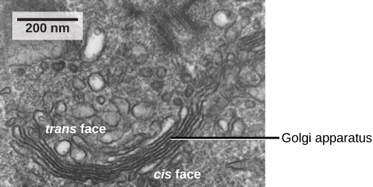
Electron micrographs below is a collection of electron micrographs with labelled subcellular structures that you should be able to identify. Make your work easier by using a label. You are expected to be able to identify free ribosomes rough endoplasmic reticulum rer lysosomes golgi apparatus mitochondrion and nucleus on an electron micrograph of a liver cell as an.
The electron micrograph of lysosomes the electron micrograph of plastids the electron micrograph of nucleus 1. Related for label the transmission electron micrograph of the nucleus black and white water bottle labels december 17th 2017 free labels wide collections of all kinds of labels pictures online. Also be sure to observe any electron micrographs which are made available in the laboratory by the instructor.
Figure 2 a a transmission electron microscope. Transmission electron microscopy allows the direct visualization of the individual components of the cell nucleus. Transmission electron micrograph showing the nucleus of a mouse cardiac tissue with nucleolus.
The dark area of the nucleus contains densely packed dna. Unlike fluorescence microscopy which relies upon the use of fluorescent probes to tag structures tem is capable of visualizing the structures themselves. The nucleus and nucleolus section 43 mitochondria section 410 and golgi apparatus section 47 can be seen.
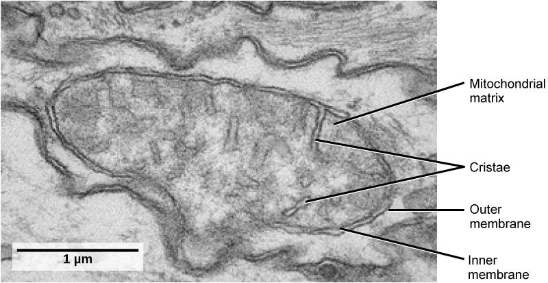
B a transmission electron micrograph of a frog leukocyte white blood cell. Nuclear envelope nucleolus nucleus heterochromatin reset zoom. It is an electron micrograph of cells largest and most important organelle the mitochondria and is characterized by the following features fig.
![]() Transmission Electron Micrographs Of Mesophyll Cells From
Transmission Electron Micrographs Of Mesophyll Cells From
 Combining High Pressure Freezing With Pre Embedding Immunogold
 An Overview Of Cellular Ultrastructure In Benthic Foraminifera
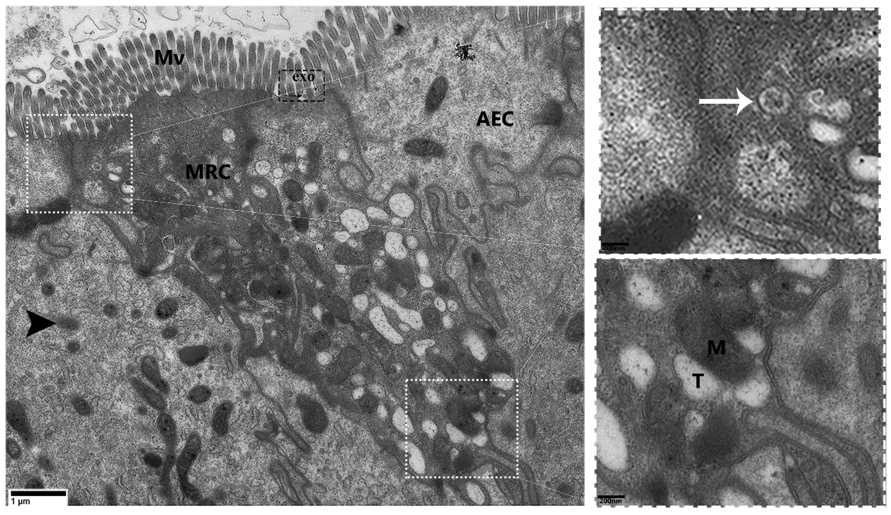 Animals Free Full Text Mitochondria Rich Cells A Novel Type
![]() Transmission Electron Microscopy Images Stock Photos Amp Vectors
Transmission Electron Microscopy Images Stock Photos Amp Vectors
![]() Immunogold Labelling Of Tail Proteins Gp45 And Gp54 A B
Immunogold Labelling Of Tail Proteins Gp45 And Gp54 A B
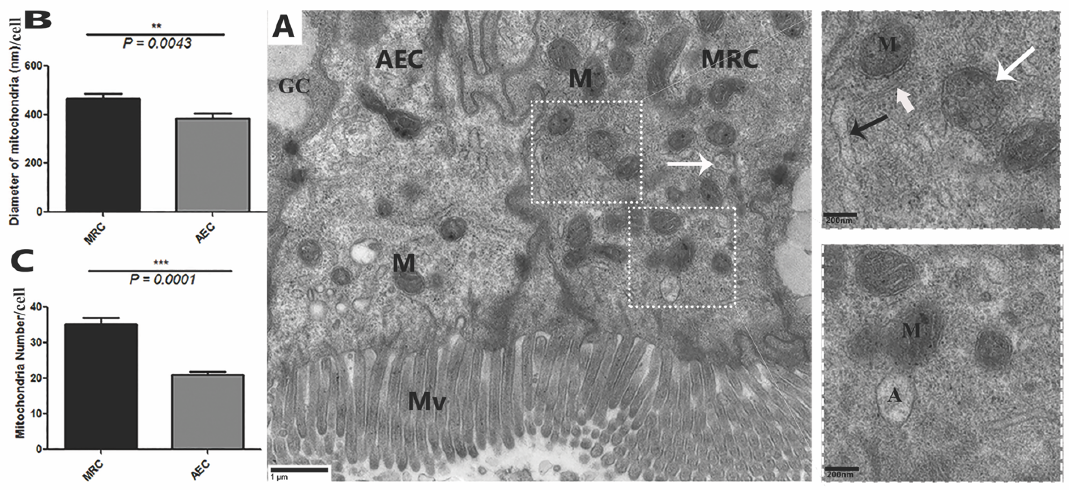 Animals Free Full Text Mitochondria Rich Cells A Novel Type
Correlative Fluorescence And Scanning Electron Microscopy Of
 Combining High Pressure Freezing With Pre Embedding Immunogold
![]() Transmission Electron Micrograph Images Stock Photos Amp Vectors
Transmission Electron Micrograph Images Stock Photos Amp Vectors
Plos One Magnetic Cell Labeling Of Primary And Stem Cell Derived
 Mitochondria And Endoplasmic Reticulum Imaging By Correlative
![]() Transmission Electron Microscopy Dna Sequencing Wikipedia
Transmission Electron Microscopy Dna Sequencing Wikipedia
 Carboxymethyl Cellulose Coated Magnetic Nanoparticles Transport
![]() Cell Organelles Cells The Basic Units Of Life Siyavula
Cell Organelles Cells The Basic Units Of Life Siyavula
 Condensins Exert Force On Chromatin Nuclear Envelope Tethers To
 A Tour Of The Cell View As Single Page
![]() Bonner Zoologische Monographien Zoology 39 Fig 20 Transmission
Bonner Zoologische Monographien Zoology 39 Fig 20 Transmission
Automated Annotating Label In Nanotomy Imaging Amp Microscopy
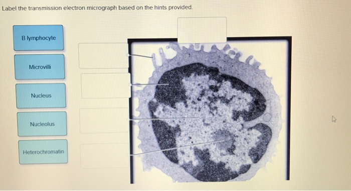 Solved Label The Transmission Electron Micrograph Based O
![]() Immunogold Labeling Of Glutathione Transmission Electron
Immunogold Labeling Of Glutathione Transmission Electron
Transmission Electron Micrograph Of Euglenid Nucleus Magnification
 1 2 Skill Interpretation Of Electron Micrographs Youtube
 Bioknowledgy 1 2 Ultrastructure Of Cells
 The Architecture Of Cell Differentiation In Choanoflagellates And
 Plunge Freezing A Tool For The Ultrastructural And


 Figure 2 From Correlated Light Serial Scanning Electron Microscopy
Post a Comment for "33 Label The Transmission Electron Micrograph Of The Nucleus"