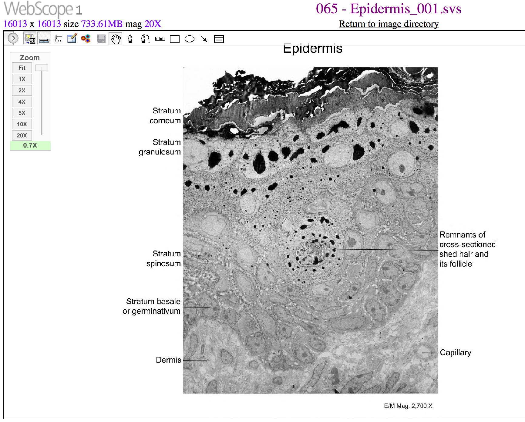
32 Label The Skin Structures And Areas Indicated
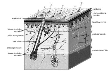
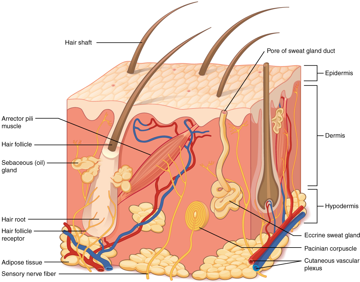
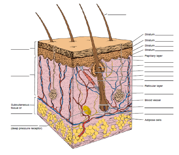
Stratum stratum layers dermal papillae séßkcedus reticular layer blood vessel hair bulb nerve fiber adipose cells 4 label the skin structures and areas indicated in the accompanying diagram of skin. Papillae hatr root sebaceous gland hal r lice arrector pili muscle reticular layer sweat gland blood vessel.
 Chromhidrosis Wikiwand
Then complete the statements that follow hair shaft stratum corneum granulosum stratum stratum spinosum stratum basale layers papillary layer denna.
Label the skin structures and areas indicated. Label the skin structures and areas indicated by leader lines and brackets on the figure. Then complete the statements that follow. Name lab time date review sheet the integumentary system 4 label the skin structures and areas indicated in the ac panying diagram of thin skin then plete the statements that follow a granules extruded wbdg the gateway to up to date information on integrated whole building design techniques and technologies the goal of whole building design is to.
Learn vocabulary terms and more with flashcards games and other study tools. Then complete the statement follow stratum stratum stratum stratum papilary layer reticular layer blood vessel tissue or adipose cells deep pressure receptor granules contain glycolipids that prevent water loss from the skin. Glands that respond to rising androgen levels are the glands.
Start studying skin structure labeling. Fibers in the dermis are produced by fibroblasts. It is located in the epidermis and the dermis.
Label the skin structures and areas indicated in the accompanying diagram of thin skin. Lamellar granules contain glycolipids that prevent water loss from the skin. Label the skin structures and areas indicated in the accompanying diagram of thin skin.
Granules extruded from the keratinocytes prevent water loss by diffusion through the epidermis. Arteries bring oxygenated blood from the heart and lungs. Select different colors for the structures below and color the coding circles and the corre sponding structures on the figures arrector pili muscle.
Granules extruded from the keratinocytes prevent water loss by diffusion through the epidermis. Veins return oxygen depleted blood back to the heart and lungs. Fibers in the dermis are produced by.
Then complete the statements that follow. Nerve fibers 0 adipose tissue 0 sweat sudoriferous gland c sebaceous gland at hair follicle 1. The hair is nourished by the follicle at its base this is also where the hair grows.
Read the definitions then label the skin anatomy diagram below. Label the skin structures and areas indicated in the accompanying diagram of thin skin. Label the skm structures and areas indicated in the accompanying diagram of thin skin.
Fibers in the dermis are produced by. Label the skin structures and areas indicated in the accompanying diagram of thin skin. Then complete the statements that follow.
64 review sheet 6 lact stratum stratum stratum.
 5 1 Layers Of The Skin Anatomy And Physiology
 Modes Of Cardiac Pacing Nomenclature Selection And Indications
 Transfer Learning With Deep Convolutional Neural Network For
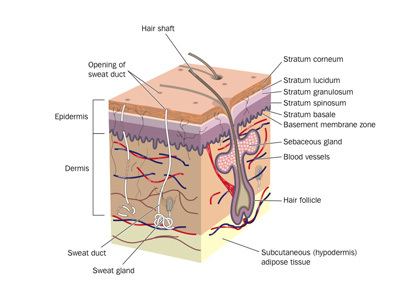 Structure And Function Of The Skin Wound Care Education Clinimed
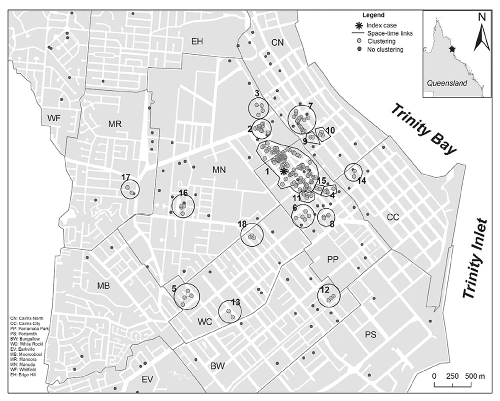
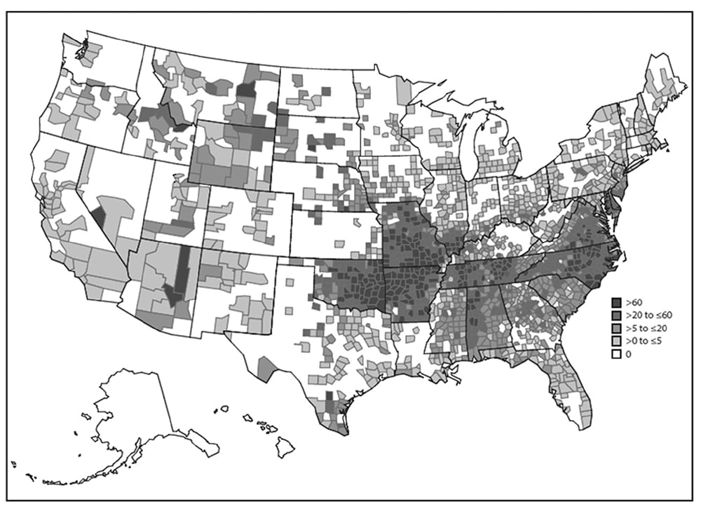 Describing Epidemiologic Data Epidemic Intelligence Service Cdc
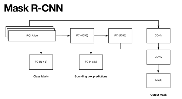 Keras Mask R Cnn Pyimagesearch
 Selfish Mutations Dysregulating Ras Mapk Signaling Are Pervasive
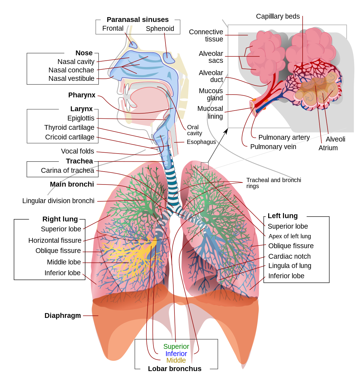 Respiratory System Wikipedia

Classification Of Circulating Tumor Cells By Epithelial
 Reproductive System The Pig Site
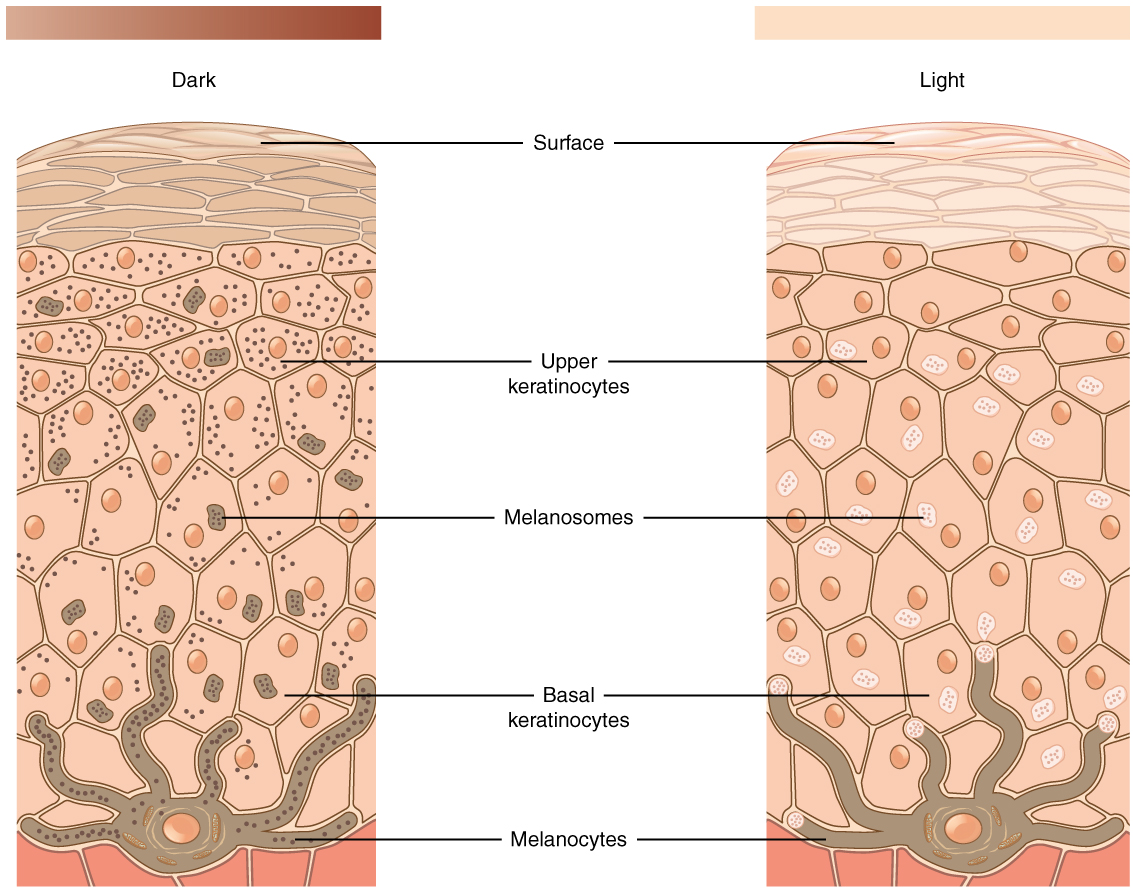
 Skin Anatomy Overview Epidermis Dermis
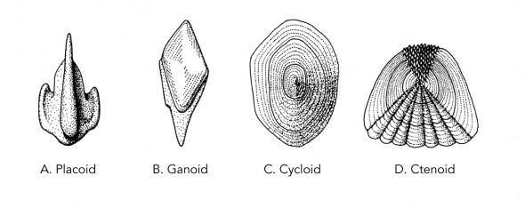 Structure And Function Fish Manoa Hawaii Edu
 5 1 Layers Of The Skin Anatomy And Physiology
 5 1 Layers Of The Skin Anatomy And Physiology
 Solved Y Structures And Areas Indicated In The Diagram 4
 A Diagrammatic Representation Of The Structure Of Human Skin In
 Introductory Anatomy And Physiology Text Amp Lab Manual Lab
 Integumentary System Development Embryology
Type Material Comparison Of Possible Cryptic Species Of The Genus
 Video Anatomy Of A Skin A3 Sylvia S H810 Online Resource
 How To Remove Your Tattoo From Home The Natural Way Skin
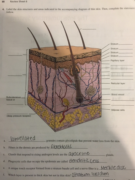 Solved 88 Review Sheet 6 4 Label The Skin Structures And
 Solved Label The Skin Structures And Areas Indicated In The Ac
.ashx?h=283&la=en&mh=360&mw=520&w=220&hash=283EF63848EC1821C83DEE53032076E7D5F51E95)
 Describing Epidemiologic Data Epidemic Intelligence Service Cdc
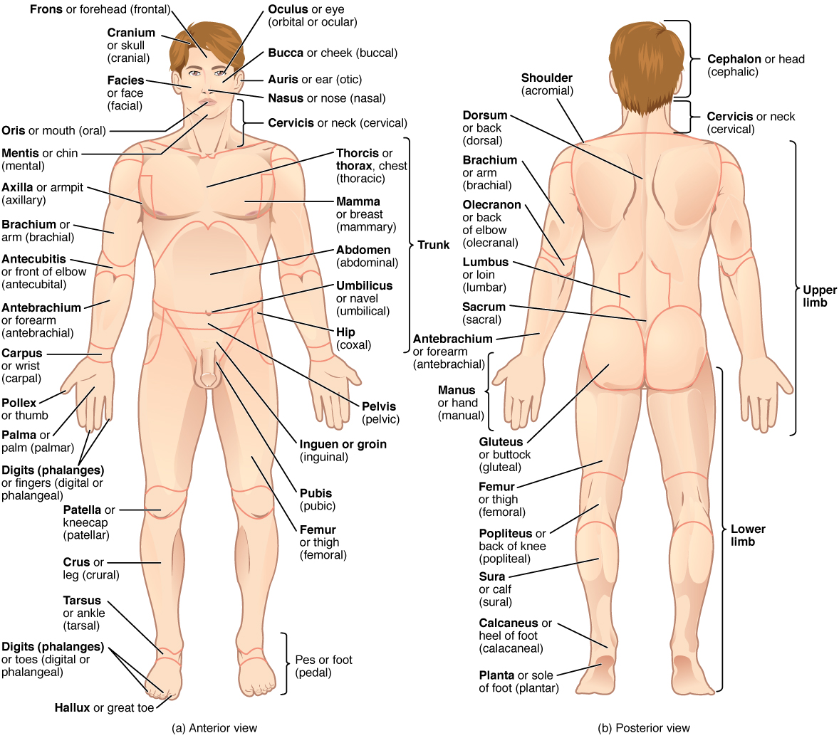 1 6 Anatomical Terminology Anatomy And Physiology
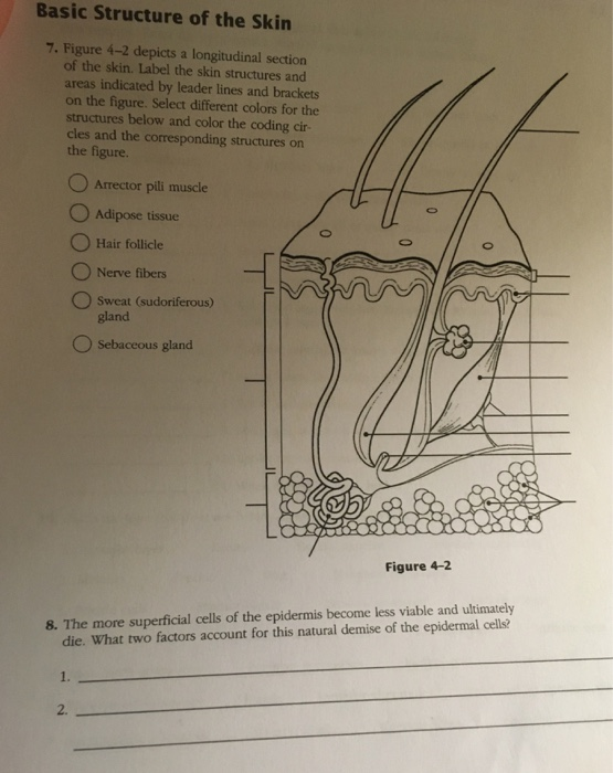 Solved Basic Structure Of The Skin 7 Figure 4 2 Depicts
 Skin Wikipedia
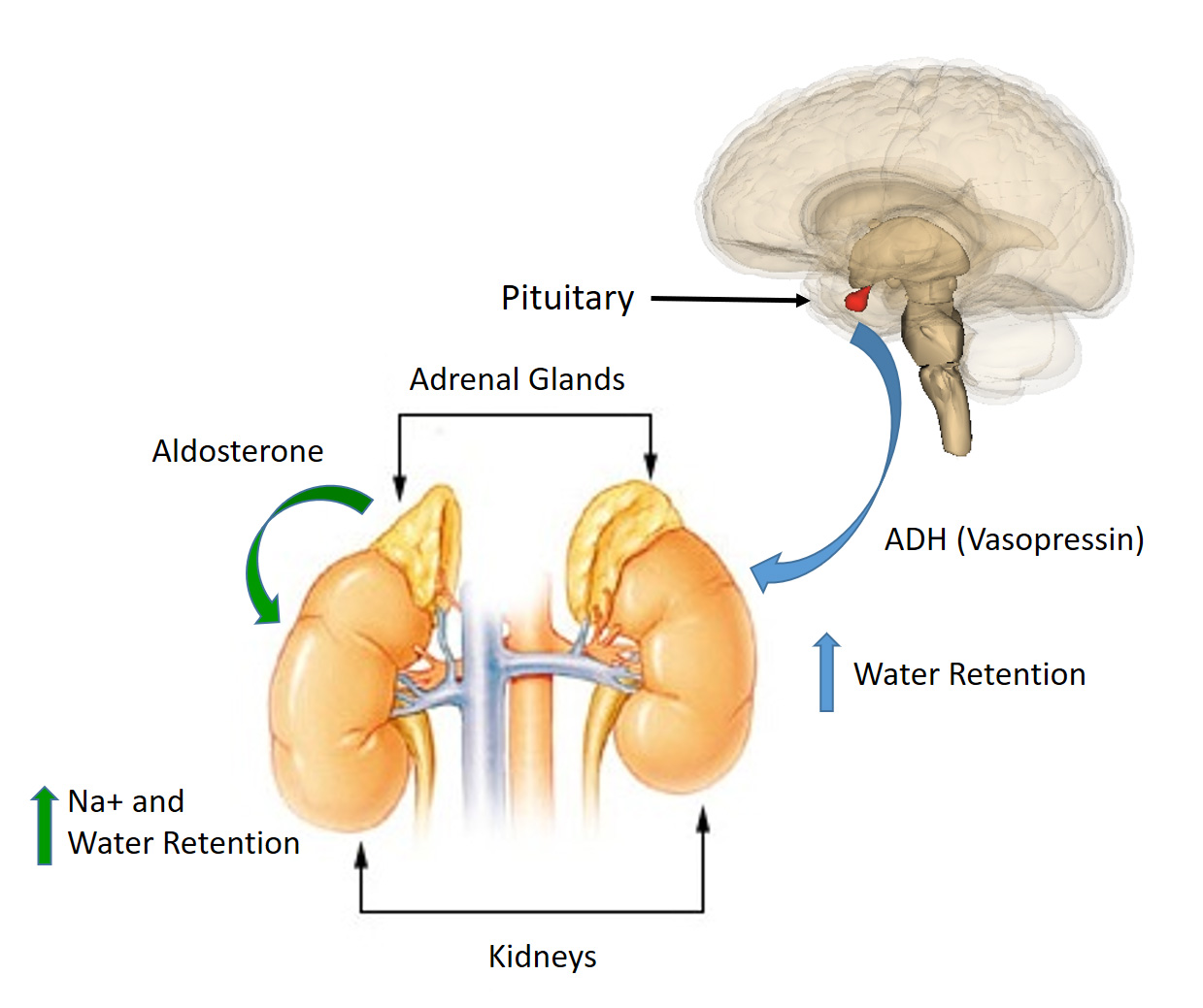
Ch103 Chapter 8 Homeostasis And Cellular Function Chemistry
 Diagram Of Longitudinal Section Of The Cow S Udder Illustrating
Post a Comment for "32 Label The Skin Structures And Areas Indicated"