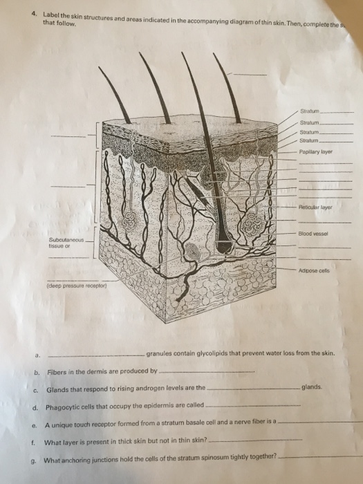
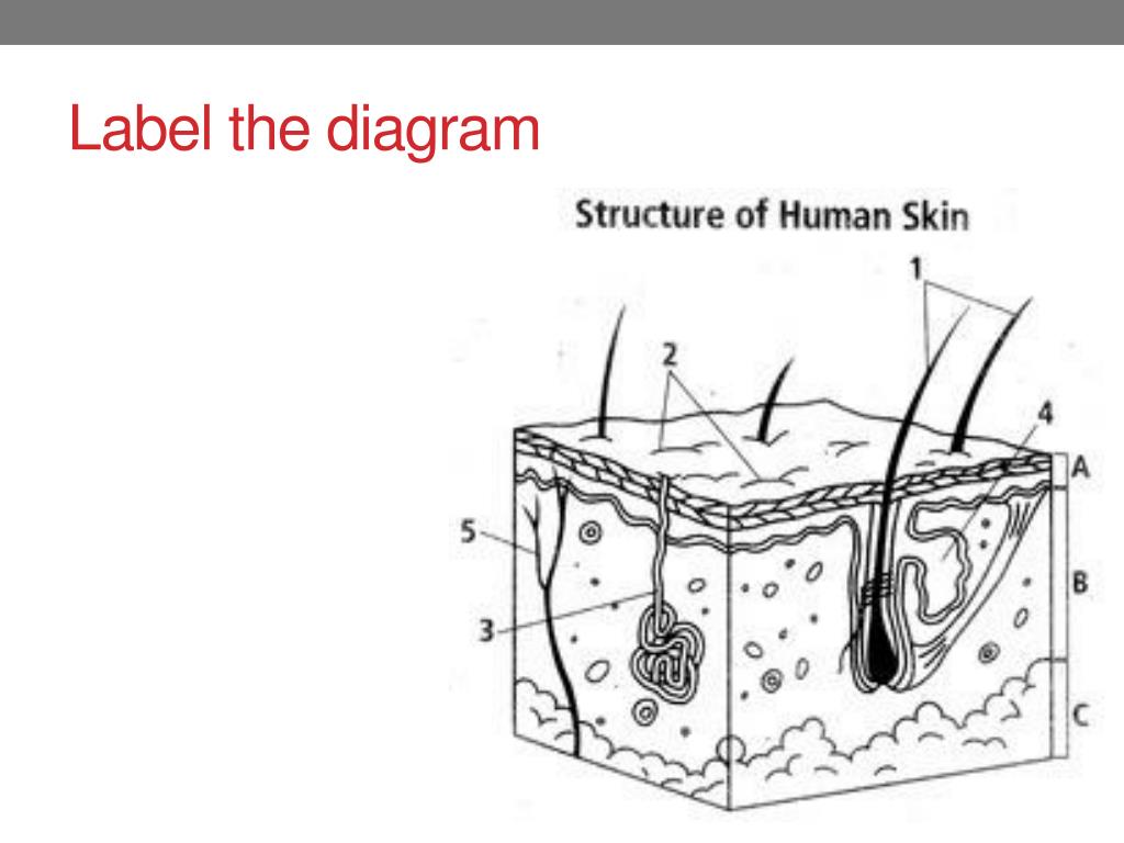
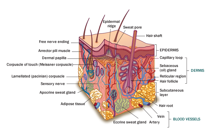
30 Label The Structure Of The Skin
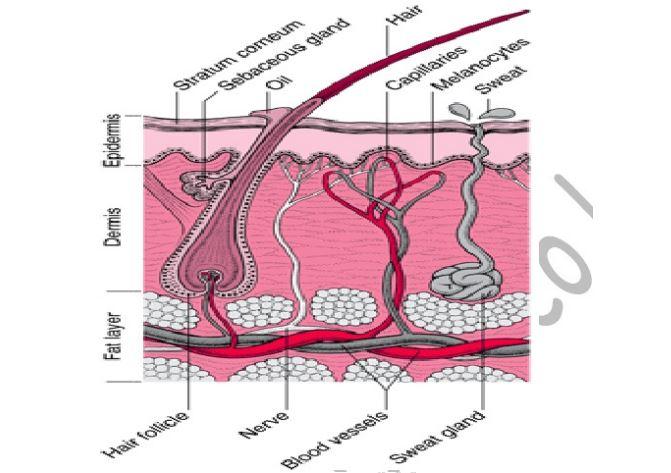
The epidermis the epidermis is the outer layer of the skin and is formed of five sub layers. Then complete the statements that follow.
 5 1 Layers Of The Skin Anatomy And Physiology
Anatomy the anatomy of the integumentary system.

Label the structure of the skin. Glands that respond to rising androgen levels are the glands. Start studying skin structure labeling. The epidermis is a tough coating formed from overlapping layers of dead skin cells.
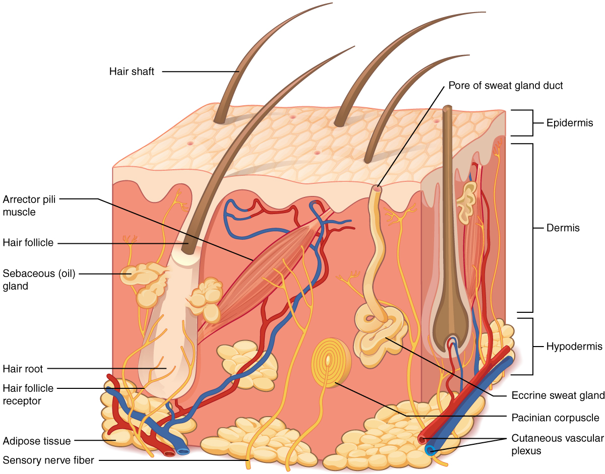
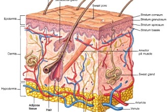
The skin is the largest organ of the body with a total area of about 20 square feet. Test your knowledge on this science quiz to see how you do and compare your score to others. Skin structure objectives students will be able to name the layers of the skin understand the structure of the skin and be able to label it from the outer surface inward.
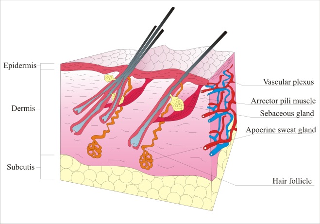
Fibers in the dermis are produced by. Granules extruded from the keratinocytes prevent water loss by diffusion through the epidermis. Label the structures of the skin and subcutaneous tissues.
Ati on gy ue ls s el s e c t. The skin protects us from microbes and the elements helps regulate body temperature and permits the sensations of touch heat and cold. Learn vocabulary terms and more with flashcards games and other study tools.
Diagram of human skin structure. The epidermis acts as a barrier between our internal body parts and the external environment. What are the names and functions of the two main layers of the skin.
Time frame to complete 30 minutes nrs efl 4 ert. A x x x standards addressed in lesson read with understanding. The outermost layer is termed as the epidermis which is made up of numerous cells called keratinocytes.
Concerning the structure of skin it is comprised of three main layers. Ardio ng n h imt mt c ng ther. Its easy to take your skin for granted but when you consider how it protects your body from harm it is something we should appreciate more.
General physiology questions on human physiology. Can you identify the skin structures in the diagram. Label the skin structures and areas indicated in the accompanying diagram of thin skin.
From outer to inner the layers are named the.
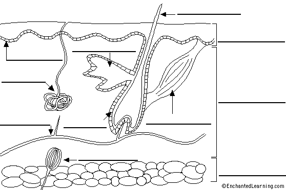 Label Skin Diagram Printout Enchantedlearning Com
 Skin Wikipedia
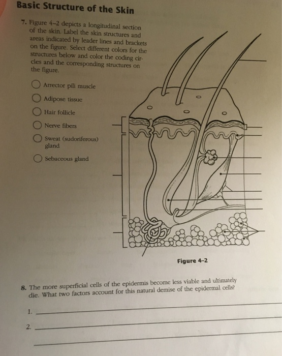 Solved Basic Structure Of The Skin 7 Figure 4 2 Depicts
 Anatomy Gross Anatomy Physiology Cells Cytology Cell Physiology
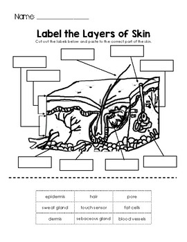 Layers Of Skin Worksheets Amp Teaching Resources Tpt
 The Integumentary System Lesson 0384 Tqa Explorer
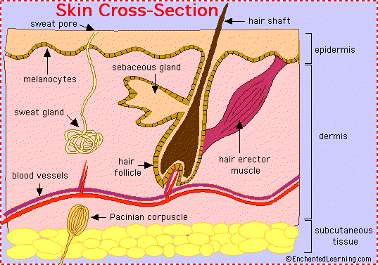 Skin Anatomy Enchantedlearning Com

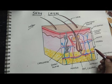 How To Draw Integumentary System Skin Layers
 A Skin Structure Quiz Proprofs Quiz
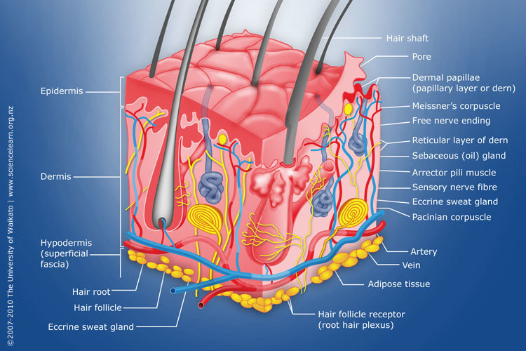 Diagram Of Human Skin Structure Science Learning Hub
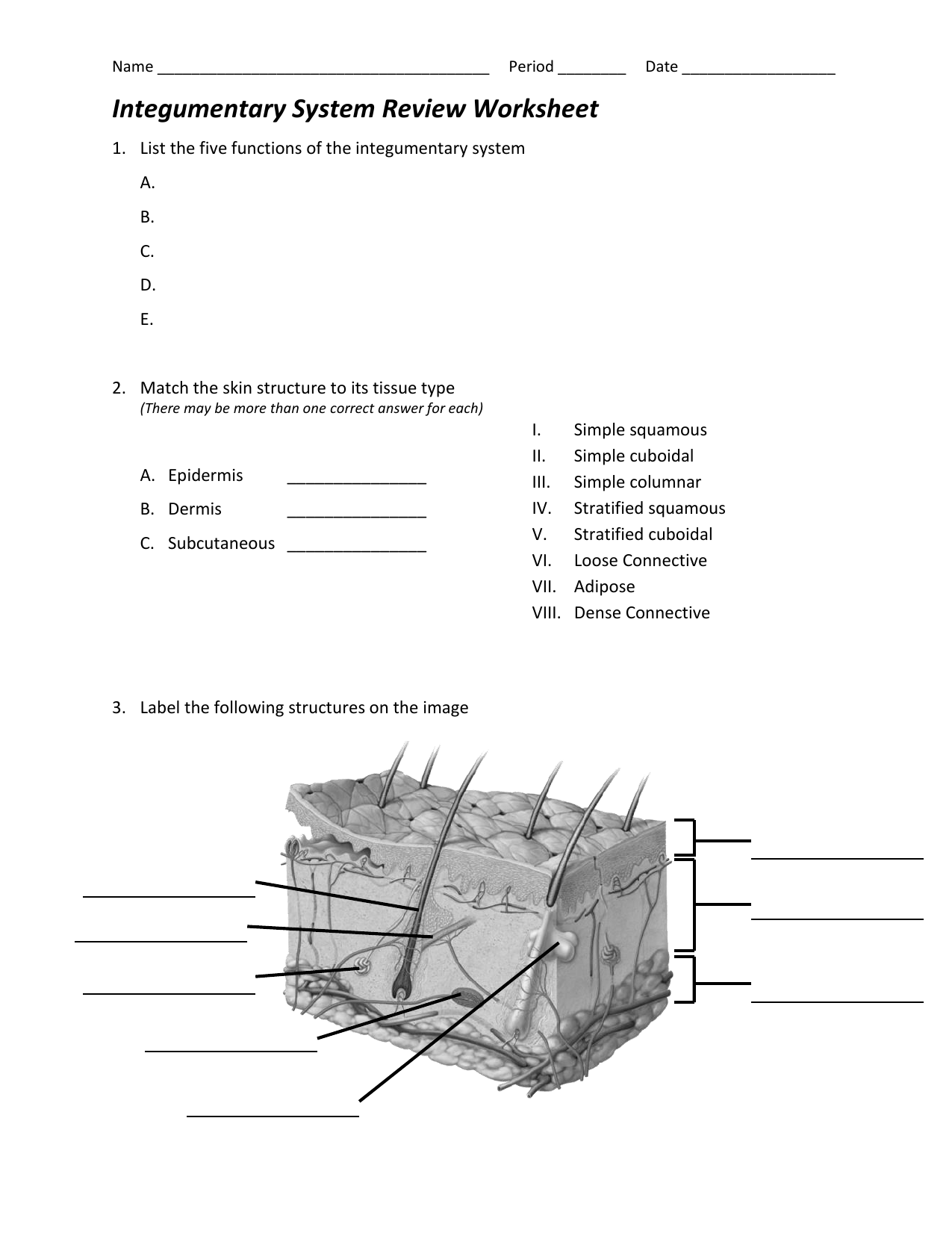 Integumentary System Review Worksheet
 Foundations Histology Cells And Tissues Embryology
 The 5 Layers Of Your Skin Dr Leslie Baumann
 The Integumentary System Lesson 0384 Tqa Explorer

 File Skin Anatomy Jpg Wikimedia Commons
 Mammary Gland Anatomy Britannica
 Skin Label Stock Illustration Illustration Of Drawing 46887482
 Structure Of Skin Biological Science Picture Directory
 Ppt Skin Appendages Hair Nails Glands Powerpoint Presentation
 Draw And Label A Mammalian Skin
 Dermatology In Dogs And Cats Intechopen
 Layers Of Skin Skincare Face Treatments Ent Wellbeing Sydney
 Stratum Basale Stock Illustrations Images Amp Vectors Shutterstock
 Label The Skin Anatomy Diagram Tag Human Skin Diagram Label Human

Post a Comment for "30 Label The Structure Of The Skin"