35 Label The Transmission Electron Microscope Image Of A Chloroplast Below
Scanning and transmission electron micrographs of chloroplasts at the full yellow stage of broccoli floret sepals durin g storage at 20 c. In the preparation of specimens for sem sepals of broccoli florets were fractured in liquid nitrogen with dmso after fixation with oso 4 as described in the text.
 The Plastid Envelope Chloroplast Manganese Transporter1 Is
Electron microscopy is the one with the most.
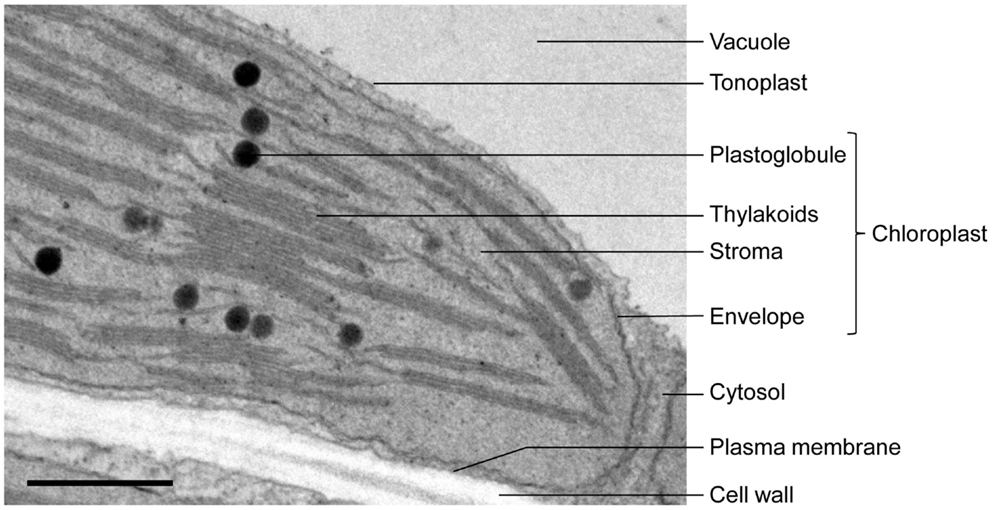
Label the transmission electron microscope image of a chloroplast below. Blc b explain why chlorophyll is found there. Perform a screen capture of the chloroplast then label a thylakoid a granum the stroma and the outer chloroplast membrane. Explain how the interrel structure of chloroplasts helps absorb the maximum amount of light.
Ocular body nose piece objective lens mechanical stage aperture diaphragm base field diaphragm right top bottom. Grana of thylakoids and their connecting lamellae are clearly visible. Sample preparation techniques and image information núria cortadellas almudena garcia and eva fernández unitat de microscòpia electrònica ccitub universitat de barcelona.
Chloroplasts are relatively large organelles with a diameter of 5 μm and width of 25 μm and so can be readily analysed by electron microscopy. Each chloroplast is enclosed by two envelope membranes which encompass an aqueous matrix the stroma and the thylakoids. ûesctbü where chlorophyll is found in a chloroplast.
Transmission electron microscope image of a chloroplast. Use a compound light microscope to view chloroplast some bacteria plant and animal cells human egg fish egg frog egg anabaena use a stereo microscope to view. The role of pigments and light.
Transmission electron microscopy in cell biology. In land plants chloroplasts are generally lens shaped 310 μm in diameter and 13 μm thick. Tem image of a single chloroplast label the transmission electron microscope image of a chloroplast below.
Arm coarse focus fine focus stage adjustment. Chloroplast 97 results electron micrograph of a thin section through a portion of the chloroplast of the unicellular green alga chlamydomonas. Left top bottom.
Transmission electron microscope image of a thin section cut from begonia spp. The micrograph is displayed as if using a virtual electron microscope so you will need to magnify the image and move to a region that contains the clearest view of chloroplast internal structures. Free ribosomes are seen as dark particles within the chloroplast stroma.
Submit your labeled image to webassign for question 2. Leaf showing a chloroplast close up view of a cell wall biology background tobacco nicotiana benthamiana leaf hairs fluorescence microscopy objective lens 20x. Label the image to review the components of a compound light microscope.
 Openstax Biology Ch4 Cell Structure Top Hat
 A Tour Of The Cell View As Single Page
 Frontiers When Proteomics Reveals Unsuspected Roles The
 Difference Between Plant And Animal Cells Cells As The Basic
 Transmission Electron Micrographs Of Armed Chlorenchyma Cells Of O
 Chlorophyll Catabolism Precedes Changes In Chloroplast Structure
Chloroplast Translation Structural And Functional Organization
 Lefkothea Regulates Nuclear And Chloroplast Mrna Splicing In
 Endoplasmic Reticulum Remodeling Induced By Wheat Yellow Mosaic
The Metabolite Pathway Between Bundle Sheath And Mesophyll
 The Molecular Biology Of Plant Cells
 Response Of Mature Developing And Senescing Chloroplasts To
Knocking Down Of Isoprene Emission Modifies The Lipid Matrix Of
 File Chloroplast In Leaf Of Anemone Sp Tem 30000x Png Wikimedia
 Transmission Electron Micrograph Showing The Immunogold Labeling
 A Tour Of The Cell View As Single Page
Moss Chloroplasts Are Surrounded By A Peptidoglycan Wall
Chloroplast Translation Structural And Functional Organization
 Cell Organelles Cells The Basic Units Of Life Siyavula
 File Chloroplast In Leaf Of Anemone Sp Tem 85000x Png Wikimedia
 Electron Microscopy Springerlink
Photosystem Biogenesis Is Localized To The Translation Zone In The
 Chloroplast Biogenesis Control Of Plastid Development Protein
Revisiting The Algal Chloroplast Lipid Droplet The Absence Of
 1 2 Skill Interpretation Of Electron Micrographs Youtube
Revisiting The Algal Chloroplast Lipid Droplet The Absence Of
 Maize Defective Kernel5 Is A Bacterial Tamb Homolog Required For
 Studying The Supramolecular Organization Of Photosynthetic
 Microorganisms Free Full Text Probing The Role Of The
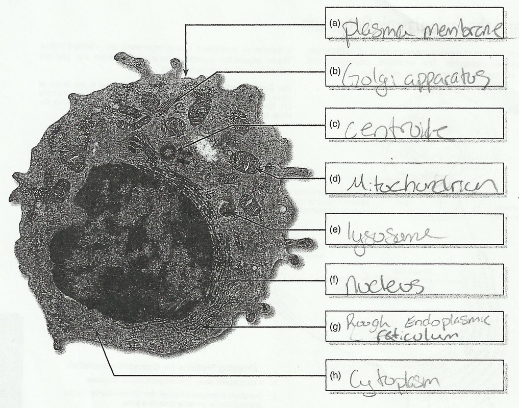 Bookfanatic89 Diagram Of Plant Cell Under Electron Microscope
Initial Events During The Evolution Of C4 Photosynthesis In C3


Post a Comment for "35 Label The Transmission Electron Microscope Image Of A Chloroplast Below"