31 Label The Tissues And Structures In This Micrograph
Label the number and structures. Label the intestinal epithelial cell in the light micrograph.
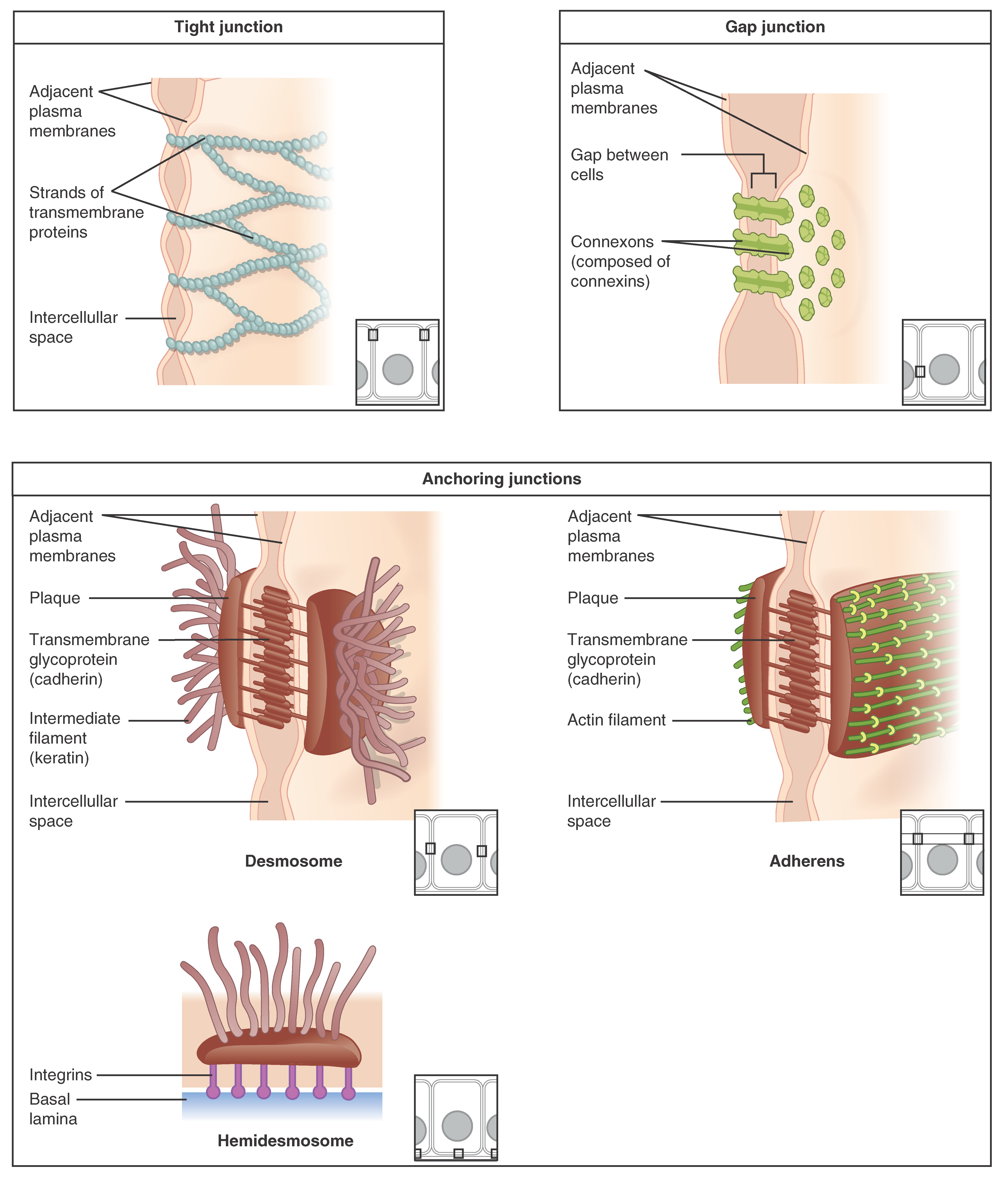 4 2 Epithelial Tissue Anatomy And Physiology
This tissue has three basic components.

Label the tissues and structures in this micrograph. Epithelial tissue connective tissue muscle tissue and nervous tissue. Layer of dividing cells 4. Connective tissue is the most diverse and abundant type of tissue.
Label the number and structures. Label the number and structures. In order to view these tissues samples were taken from organs.
Label the light micrograph of the seminiferous tubule using the hints provided. Stratified squamous epithelium 1surface of tissue 2. Label the tissues and structures in this micrograph.
Identify the structure indicated. 14 nucleus of columnar epithelial cell simple columnar epitheliunm ciliated skipped lamina propria lumen of uterine tube columnar epithelial cell ciliated membrane stratified columnar epithelium ciliated reset zoom. It is characterized by a much larger content of extracellular material matrix than other tissues.
Categorized into one of four groups. Complete the table identifying the source and target tissue of each of the female sex hormones. Label the number and structures.
Label the structures associated with blood and bile flow through the hepatic lobule. Label the number and structures. Test your knowledge on this science quiz to see how you do and compare your score to others.
Note that the structures in the right half of the pelvis have been retracted. Label the structures seen in this superior view of the female pelvis. Compact bone osseous tissue central canal.
Learn more about adipocytes and types of adipose tissue at kenhub. The disorder of the large intestine producing a cobblestone effect within the tissues of the colon is. Were describing the structure and function of the adipose connective tissue.
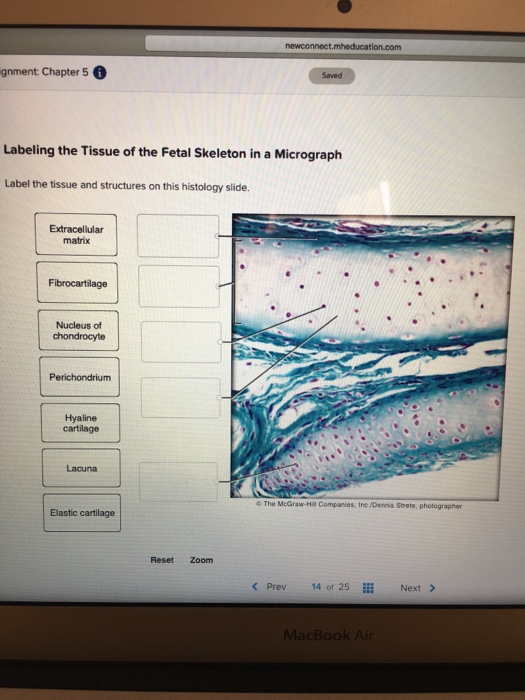
Stratified cuboidal capillary lacuna of bladder umbrella cell transitional lumen of bladder dense connective tissue loose connective tissue reset zoom prev 22 of 25 next. Chapter 5 labeling the tissue of the urinary bladder in a micrograph label the the tissues and structures on the histology slide. Type the name of the tissue indicated by the question mark.
Cells of the blood. Identify the tissue type and a location where it is found. It is designed to support protect and bind organs.
Identify the structure indicated. Organs are macroscopic structures which are composed of more than one tissue type and perform a specific function for a multi cellular organism. Cells protein fibers and ground substance a non living material produced by the cells.
 Plant Tissues Plant And Animal Tissues Siyavula
:max_bytes(150000):strip_icc()/loose_connective_tissue-5b68c53446e0fb0050388e5a.jpg) Connective Tissue Types And Examples
Connective Tissue Types And Examples
 Functions Of Transitional Epithelium Tissue
 Structure And Function Of Skin Biology For Majors Ii
Blue Histology Epithelia And Glands
 Plant Tissues Plant And Animal Tissues Siyavula

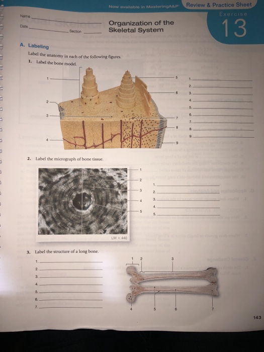
 Solved Review Amp Practice Sheet Exercise Organization Of T
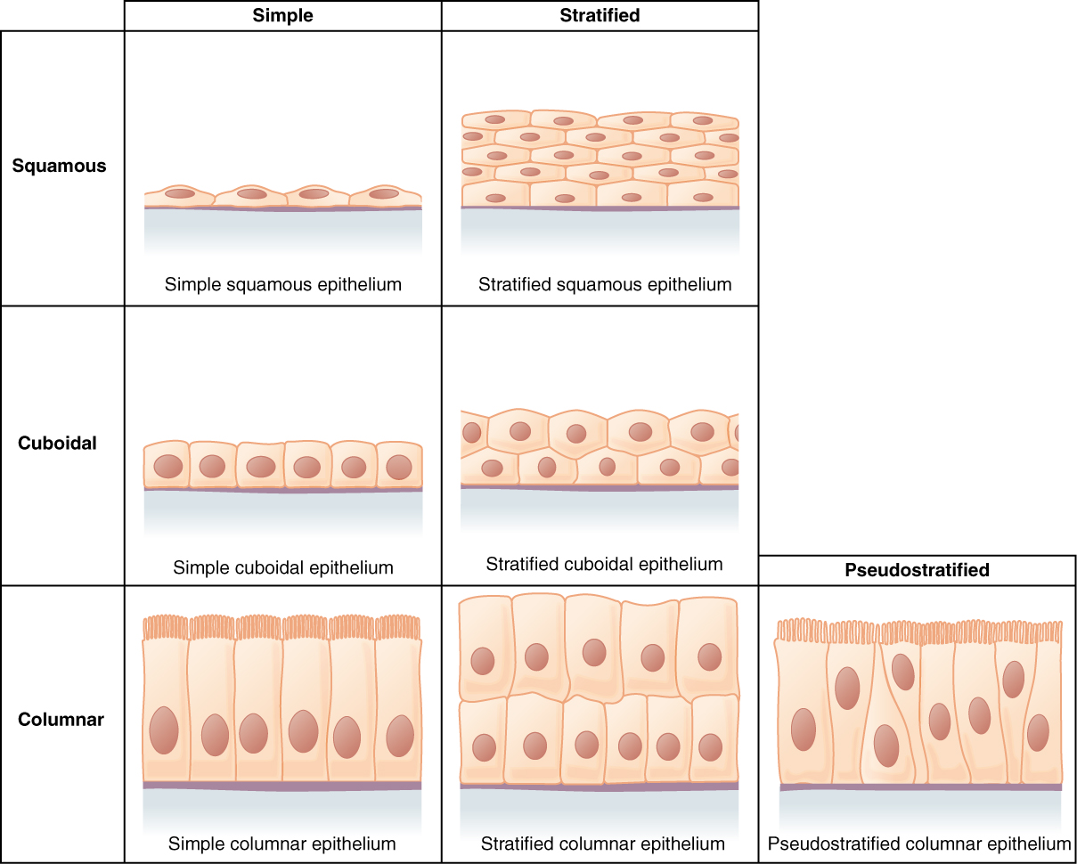 4 2 Epithelial Tissue Anatomy And Physiology
 Connective Tissue Anatomy And Physiology
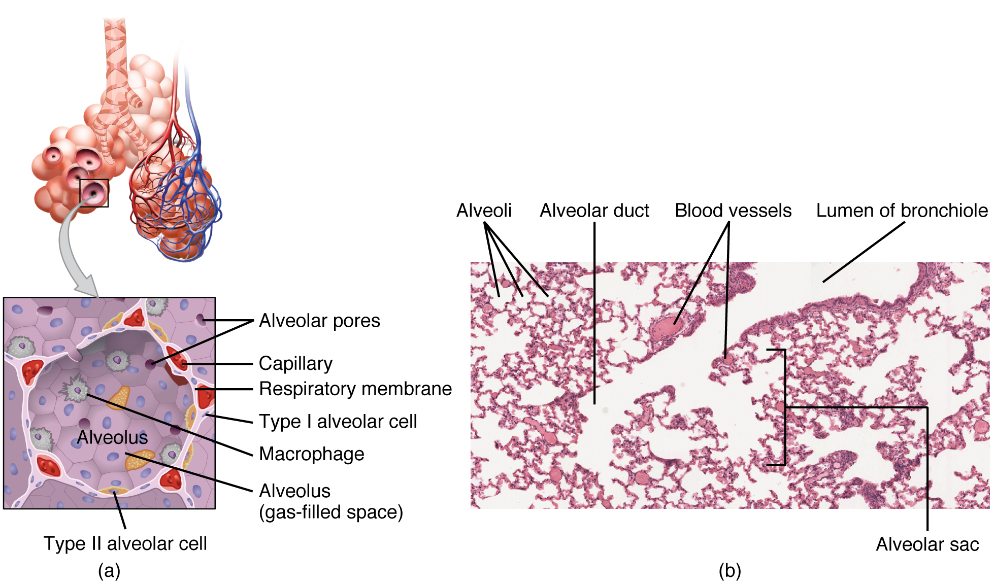 22 1 Organs And Structures Of The Respiratory System Anatomy And
 Animal Tissues Plant And Animal Tissues Siyavula
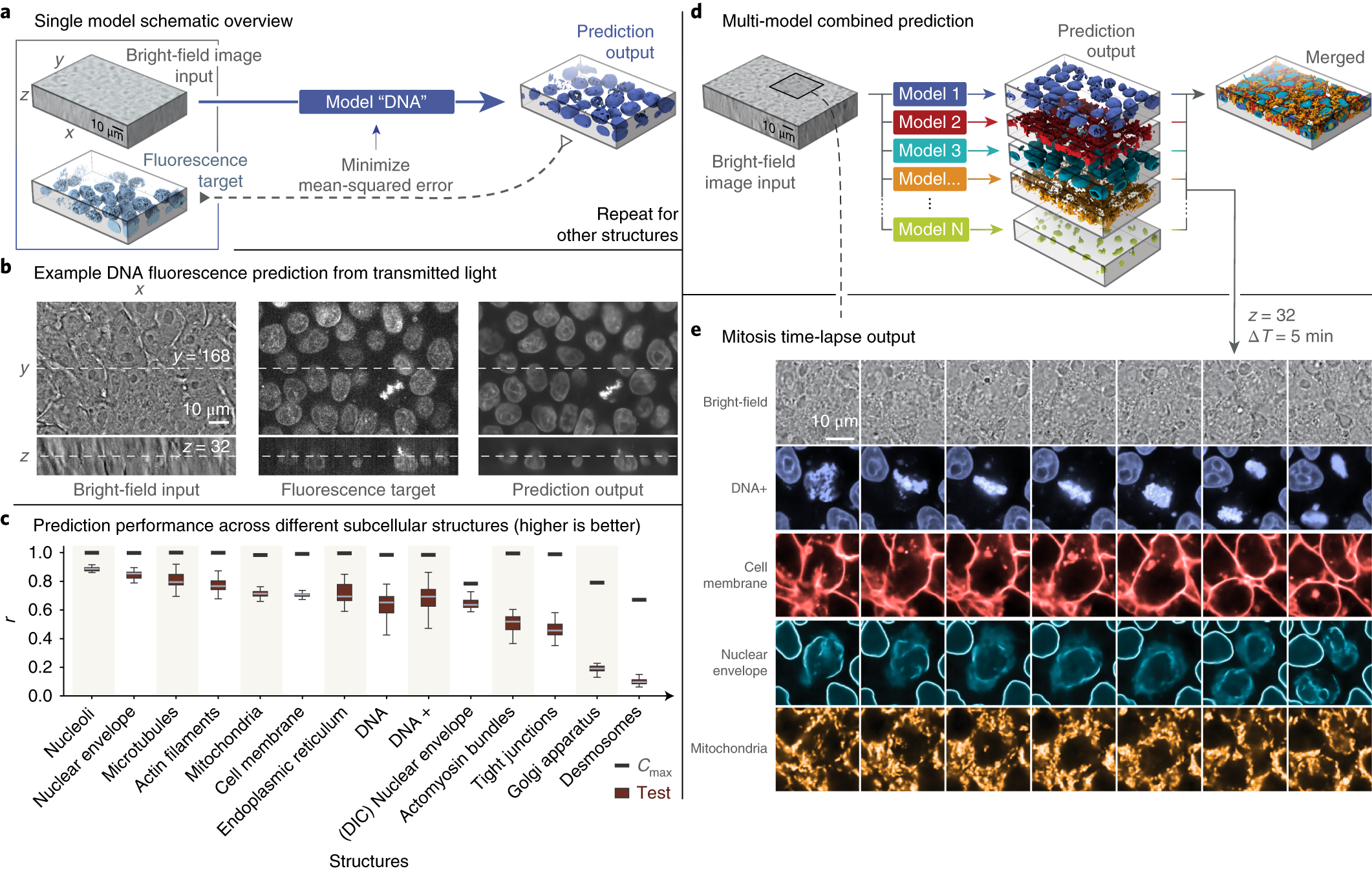 Label Free Prediction Of Three Dimensional Fluorescence Images
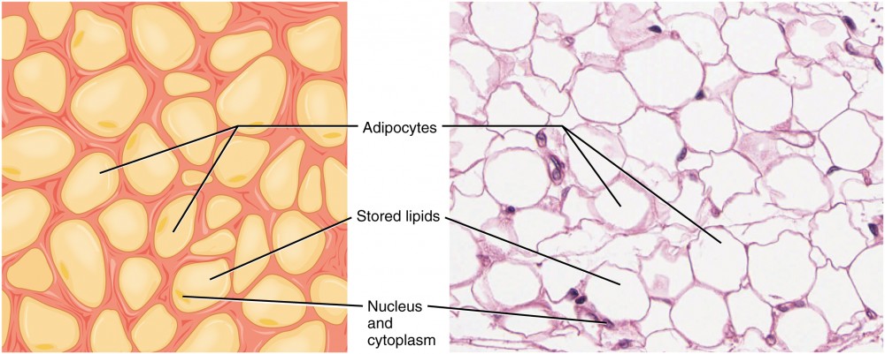 Connective Tissue Anatomy And Physiology
 Bone Tissue And Cells Under The Microscope
 Bone Tissue And Cells Under The Microscope
 Examining Epithelial Tissue Under The Microscope Human Anatomy
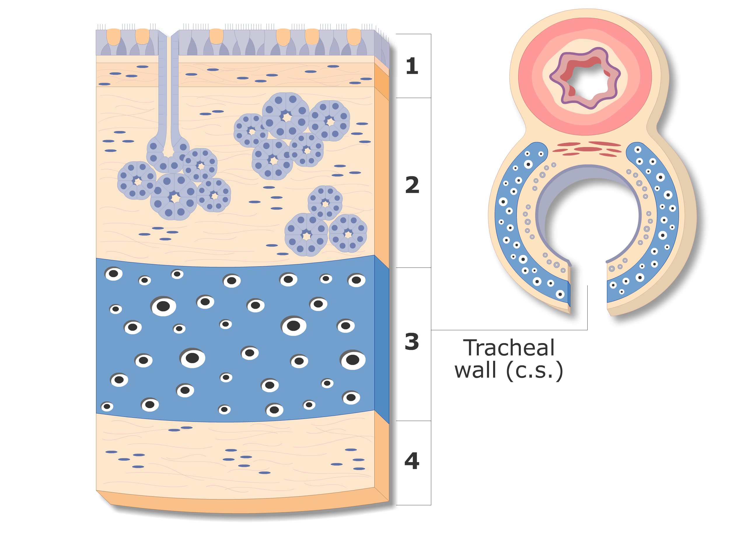 Tracheal Wall Composition And Structure Anatomy Of The Tracheal
 Animal Tissues Plant And Animal Tissues Siyavula
/types-of-cells-in-the-body-373388-v3-5b76f0ad46e0fb0050ba820e.png) 11 Different Types Of Cells In The Human Body
11 Different Types Of Cells In The Human Body
 Connective Tissue Anatomy And Physiology
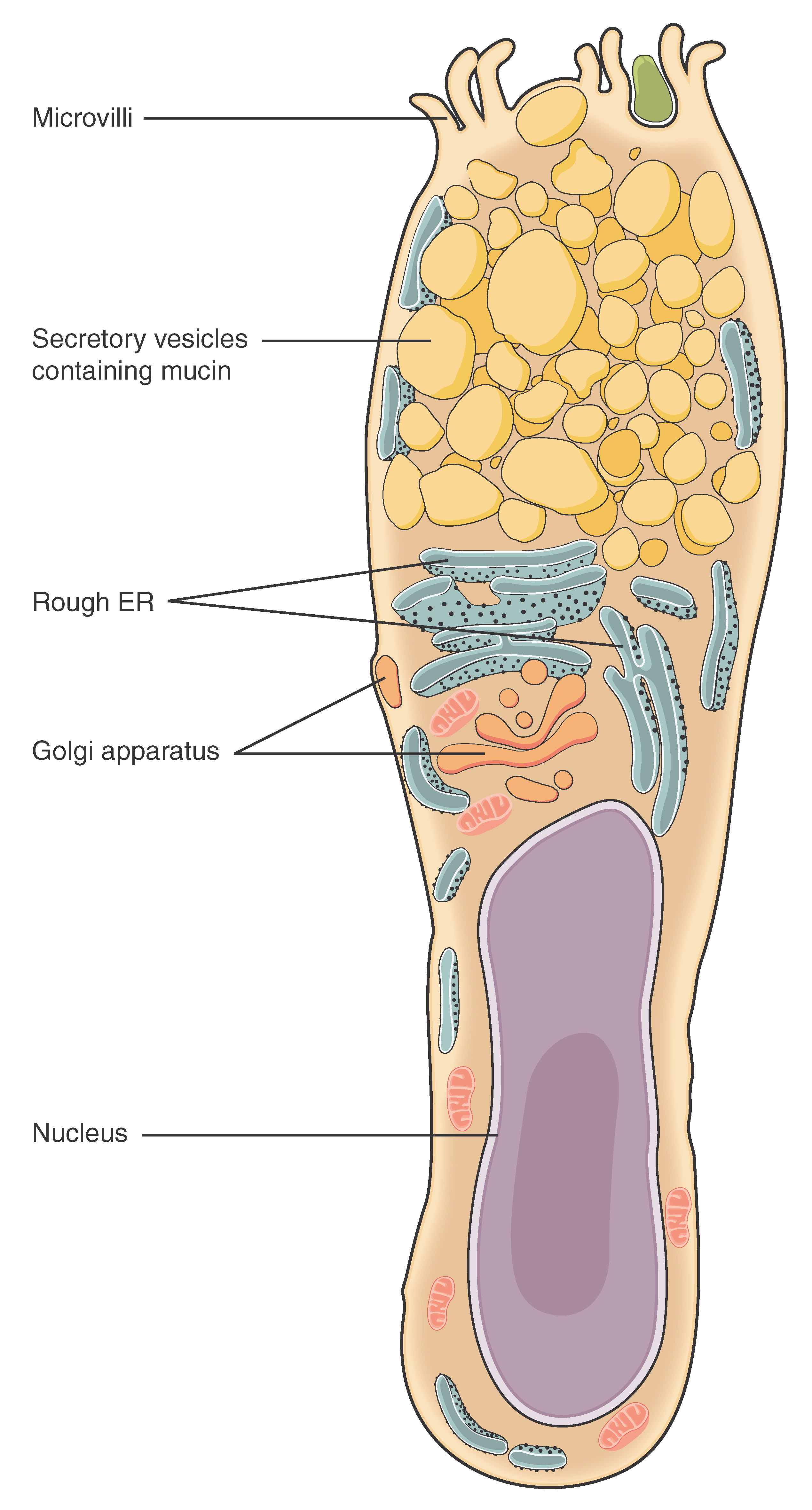 4 2 Epithelial Tissue Anatomy And Physiology


Post a Comment for "31 Label The Tissues And Structures In This Micrograph"